Tuesday, November 9, 2010

Physical Exam Tapas


Today we saw some great cases:


  • Erythema Nodosum: discussed here. Secondary to sarcoid? TB?

  • CREST syndrome: discussed here.

  • Pulmonary Hypertension: discussed here. Secondary to Ebstein's anomaly.

Tuesday, November 2, 2010

Parkinson's Disease


Today we saw a gentleman with Parkinson's Disease. A few features to watch for on the physical exam.
  • Head and Neck: Look for a masked facies, seborrhea, absence of blinking, and possibly drooling. In addition, the glabellar tap maneuver is often positive, but not a very nice exam to perform.
  • Listen: Speech is often soft, slow, and monotonous.
  • Tremor: This is at rest, and initially starts unilaterally. Often described as a 'pill rolling' tremor with 4-6 cycles per second. It will eventually move bilaterally.
  • Tone: Classically with 'cogwheel' vs lead pipe rigidity. We felt a good example of cogwheeling today.
  • Bradykinesia: Slower movements. You can bring this out by having them pretend to play the piano, heel tapping, etc.
  • Gait: Watch for a stooped posture ('simian posture'), with a shuffling ('festinating') gait. It appears as if their center of gravity is just in front of them. Watch closely when turning around - this often requires multiple little steps rather than just one or two steps.
  • Other: Watch for micrographia (small writing), difficulty opening jars, difficulty turning in bed, difficulty going from a sit-to-stand.
Here is an excellent JAMA article on the clinical evaluation of Parkinson's Disease.

Tuesday, October 26, 2010

Peripheral Stigmata of Liver Disease


1. Liver Failure - some exam findings to look for include:
  • encephalopathy
  • asterixis
  • jaundice
  • bruising
  • fetor hepaticus
  • muscle wasting
  • clubbing (read more here)

2. High Estrogen states associated with liver disease, look for:

  • palmar erythema
  • spider nevi (these blanch)
  • gynecomastia
  • feminization of body hair
  • testicular atrophy

3. For underlying clues as to the etiology of liver disease, look for:

  • duputryen's contractures - alcohol
  • keiser fleischer rings - Wilson's disease
  • 'bronzed' complexion - iron overload syndromes (hemochromatosis)
  • track marks - IV drug use, and perhaps underlying Hepatitis C.
4. Also look for signs of portal hypertension, which include:
  • ascites and pedal edema (previously posted here and here)
  • distended abdominal veins
  • caput medusa
  • splenomegaly

Below: duputrene's contracture, palmar erythema, and spider angioma


Thursday, October 14, 2010

The Argyll Roberston Pupil


(Treponema pallidum pictured left)

Today we examined a patient for possible tertiary syphilis and looked specifically for an Argyll Roberston Pupil.

Accommodation Reflex: This is intact (CN II and III). There is pupillary constriction when an object is brought closer the patients face along with convergence of the eyes.

Pupillary Constriction to Light: This NOT intact (also CN II and III). When a light is shone into a patients eyes, there is no pupillary constriction.
We also examined a patient for aortic stenosis. You can read more about this here and here.

Tuesday, October 12, 2010

Lipids and the skin

1. Xanthelasma: cholestrol deposits under the skin, typically around the eye - often called xanthelasma palpebrarum. Usually yellowish/whitish and flat.


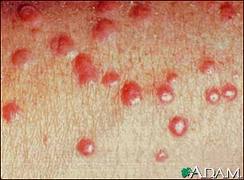
2. Eruptive xanthoma: can be pruritic, and may resolve over a few weeks. Often seen in patients with diabetes and hyperlipidemia. Pustular on an erythematous base.


3. Tendon xanthoma: classically a slowly appearing nodule on the extensor tendons of hands and feet.


4. Lipemia retinalis: Very high levels of triglycerides (>400 in the USA, >40 in the rest of the world). You can see a milky colouration to the retinal vessels.


A neat link from NEJM on lipemia retinalis over here.

Thursday, October 7, 2010

Rheumatoid Arthritis: Some hand findings

On inspection remember to look for "SEADS" - swelling, erythema, atrophy, deformities, skin changes.


Wrist:
  • Often has Radial deviation
  • You may see a 'dinner fork' deformity at the wrist

Hand:

  • Look for muscle atrophy in the interossious, thenar, and hypothenar regions
  • Look and feel for Tendon nodules, and contractures

Fingers:

  • Vasculitic nail changes: nail fold infarcts, periungual erythema
  • Ulnar deviation of MCP's
  • Swelling of the PIP's, the DIP's become involved later in the disease
  • Swan Neck deformities (see photo above)
  • Boutonniere deformitiy (see photo above)


Tuesday, October 5, 2010

Cranial Nerves III, IV, and VI


Interesting eye findings today:


Cranial Nerve III
  • All extra ocular movements except those from the Superior Oblique (CN IV) and Lateral Rectus (CN VI).
  • Innervates the Levator Palpebrae Superioris: this keeps your eyelid open. Many CN III lesions will result in ptosis.
  • Efferent limb of the pupillary light reflex. Remember to look for a "direct" and then a "consensual" response to light.
  • A Quick CN III Tip: Pupillary constriction is controlled by the outer fibers of CN III, and ocular movements/upper eyelids are controlled by the inner fibers. So lesions that compress the outer CN III fibers (eg. lesions like Posterior Communicating Artery Aneurysms) present initially with dilated pupils, while lesions affecting the inner portions of CN III (like infarction in diabetes) present with ptosis and a "down and out" position of the eye that spares the pupil.

Cranial Nerve IV

  • Innervates the Superior Oblique muscle, which moves the eye "down and in". Lesions here result in difficulty looking down. Patients have trouble walking downs stairs.

Cranial Nerve VI

  • Innervates the Lateral Rectus muscles, which moves the eye laterally.

Thursday, September 30, 2010

Tophi and Gout

Today we saw some pretty empressive gouty tophi - small joints on the hands were involved, as well as an impressive tophus on the elbow (this picture is from wikipedia, not our patient). Tophi are just uric acid crystals deposited in the tissues, and is evident usually after several years of gout. Remember to look for tophi on the joints, but also in the helix/antihelix of the ear (see picture below).


Gout:
  • Clinical: Typically presents with acute monoarthritis, occasionally a few joints involved. Red, hot, swollen, tender. Often first MTP, ankle, knee.
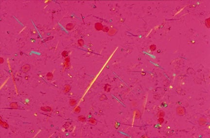
  • Evaluation: Negatively birefringent crystals visualized, often intracellular. They look like this. These are monosodium urate crystals.
  • Risk factors: hyperuricemia (lymphoma, other causes of increased cell turnover), obesity, EtOH, medications like HCTZ, asa.
  • Acute Treatment: NSAIDS, intra-articular steroids, colchicine, systemic steroids if multiple joints involved.
  • Chronic treatment: Allopurinol if multiple attacks per year (usually >2).
Pseudo Gout:
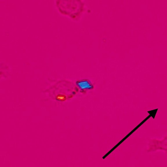
  • Rhomboid crystals, occasionally inside macrophages. They look like this. These are calcium pyrophosphate crystals.
  • Often in elderly patients with multiple medical problems.
  • Often polyarticular, and may affect wrists, shoulders.

Tophi on the ear

Tuesday, September 28, 2010

Knee Effusion

-Tunisia?
-Two Knees, Yeah!
-Two Knees, Huh?

Okay...terrible...there's plenty more where that came from. Today we examined two knees. And a few knee problems - gout, prepatellar bursitis, and septic arthritis.

Is there an effusion?

1. Inspect: in the absence of an obvious effusion, have a look at the medial side of the knee - normally there is a 'dimple' here. Expose both sides and compare. Absence of this dimple is usually indicative of an effusion.

2. Palpate: There are a few ways to feel for an effusion....
  • Patellar Tap: push on the suprapatellar region to get fluid under the patella, then press firmly on the patella. You may feel it tap on the bone underneath. Kind of uncomfortable.
  • Ballotment: push on the suprapatellar region to get fluid under the patella. Then with your other hand, use your thumb and index finger to ballot the fluid back and forth.
  • "Milk" test: 'milk' (or 'sweep') fluid from the medial side up the knee and watch that medial dimple re-appear. Then sweep the fluid back down the lateral aspect of the knee and see the medial dimple bulge out again.
  • The Forgotten 4th test: Not sure what to call this one. I learned from Dr. Mary Bell, an incredible Rheumatologist. With one hand, place your index finger and thumbs on either side of the patella. Then with your index finger of your other hand, gently press on the suprapatellar area. In the presence of an effusion, you will feel pressure on your thumb and forefinger.

Let me know if you need this explained again at the bedside - happy to show you at any time.

3. Links:


Thursday, September 23, 2010

Chest findings...."just tap it in...tap it in..."


Today we did a lot of percussion and auscultation....and a bit of tactile vocal fremitus. The summary below is very important - you can predict what you will see on chest x-ray with your history and physical exam >90% of the time. Click on it to enlarge. Here is a good link to the complete chest physical exam.


Tuesday, September 21, 2010

Splenomegaly

What does this picture have to do with splenomegaly? Send me an email - if you're right, I'll buy you a coke.

The spleen is the 3rd most underappreciated internal organ.

If you need a reminder on how to perform any of the maneuvers below, send me an email/page and we can go through it.

Does my patient have splenomegaly? Tough to say... this is not the most reliable exam, however if your pre-test probability is somewhat high (>10%), then the exam can be quite helpful.

Castell's Sign: The patient is supine. Locate the intersection between the left anterior axillary line (*note* not mid axillary line) and the last intercostal space. Perucuss. It should be resonant. When your patient breaths in and holds, this should stay resonant. If you hear dullness then voila - probably splenomegaly(Sensitivity 82%; Specificity 83%).

Palpation: Either use your hand, or the patients hand for counter-pressure in the left flank. Palpate from the RLQ diagonally up, inching incrementally as the patient exhales. Try rolling the patient into the right lateral decubitus if you do not feel anything and repeat your palpation(Sensitivity ~60%; Specificity ~90%). Feel for the splenic notch.

Middleton's Hooking Maneuver: Approach the patient from the left side, with the patient supine and their fist under the left costovertebral angle, hook your fingers under the left costal margin. Ask the patient to breath in, and feel for the spleen tip (sensitivity ~60%; Specificity ~90%).

Others: Nixon's percussion method is helpful, but Traube's space is pretty useless. I can show you these whenever you like.

Here is a link to the sentinel JAMA article on splenomegaly.

Thursday, September 16, 2010

Stevens-Johnson Syndrome

Stevens-Johnson Syndrome and Toxic Epidermal Necrolysis are about as bad a drug reaction as you can get. These are basically the same condition, with TEN being on the more serious side of the spectrum compared to SJS

SJS: Less than 10% of body surface area is affected. Mucous membranes virtually always involved. macules which are erythematous or purpuric morph into epidermal necrosis and sloughing.

TEN: See above, except >30% of the body is involved. Diffuse erythema is also seen in this condition.

Both: Associated with systemic symptoms like fever, malaise, anorexia. Bullae may form on the skin prior to sloughing.

What causes it? Mostly in response to medications. Classic drugs include allopurinol, sulfa medications, penicillins, anti-seizure drugs (dilantin, carbamazepine), and some NSAIDS.

How do your manage these cases? 1. early recognition, 2. discontinuation of offending agent, 3. supportive care, 4. wound care management, including eyes and mouth, 5. monitor for fluid/electrolyte disturbances, 6. debridement. The use of IVIG or steroids is controversial. Many patients require specialist care in a burn unit.

How does the picture above relate to this topic? send me an email...if you're right I'll give you a prize.

Tuesday, September 14, 2010

Terry's Nails and Lindsay's Nails

Today we looked at nails a bit more closely. We examined the white part of the nail, termed the Lunula, which is Latin for 'little moon'. This normally occupies a very small proportion of your nail.


Lindsay's Nails: also termed "half-and-half" nails. As the name implies, the lunula extends to roughly 50% of the nail. It is classically described in chronic kidney disease.

Terry's Nails: The lunula extends to >75% of the nail. This is described in liver diseases, CHF, diabetes, and hyperthyroidism.

Thursday, September 9, 2010

Interstitial Lung Disease

Today we saw some really interesting cases including:

*Ascites: check out a previous post with evidence-based physical exam findings here.

*Clubbing: also discussed, with evidence-based exam findings at this link.

One patient we saw had terrible interstitial lung disease. These are confusing conditions. Here's an approach as to the underlying etiology:

Systemic Diseases: ILD is found commonly in Sarcoidosis and Collegen Vascular Diseases - the prototype being Scleroderma. Remember, ILD is not so common in SLE or Rheumatoid arthritis.

Exposures: these are classically divided into organic and inorganic exposures:

  1. Organic: aka "Hypersensitiviy Pneumonitis". Think about bird fanciers lung, cheese washers lung, farmers lung, and many many more.
  2. Inorganic: aka "Pneumoconiosis". This includes dust from asbestos, silica, and coal.
Drugs: Exposure to alkylating chemotherapeutic agents (eg. bleomycin), but also commonly used drugs like amiodarone, methotrexate, nitrofurantoin, and sulfa medications are also implicated in ILD.

Idiopathic: This is a strange category with bizarre acronyms. It doesn't help that there are old and new acronyms, and that both are used frequently. The diagnosis is usually based on histology.
  1. Usual Interstitial Pneumonia (UIP), also known as Interstitial Pulmonary Fibrosis (IPF) - this is perhaps the most common idiopathic ILD.
  2. Acute Interstitial Pneumonia (AIP), also known as the 'Hamman-Rich Syndrome'. Just like IPF above, but more rapid.
  3. Cryptogenic Organising Pneumonia (COP), but if the underlying etiology is known, you will hear it called Bronchiolitis Obliterans and Organising Pneumonia (BOOP). There is lots of granulation tissue in the small airways here.
  4. Others: Desquamative Interstitial Pneumonia (DIP), Lymphocytic Interstitial Pneumonia (LIP), Non-specific Interstitial Pneumonia (NSIP)....oy vey...
Other Rare Causes of ILD: Alveolar hemorrhage, Pulmonary Alveolar Proteinosis, and my personal favorite, Pulmonary Infiltrates with Eosinophilia (PIE). mmmmmh.....pie. And Lymphangioleiomyomatosis (LAM).

Mimickers of ILD: Pneumocystis Carini Pneumonia, Lymphangitic carcinomatosis, and once in a while, good ol' congestive heart failure.

Links: A good review of Idiopathic Pulmonary Fibrosis can be found at this link.

Tuesday, September 7, 2010

Clubbing


Common causes of Clubbing:
  • Gastrointestinal: liver disease, inflammatory bowel diseases, celiac disease, malignancy
  • Pulmonary: malignancy, suppurative (empyema, abscess), Cystic Fibrosis, TB, bronchiectasis, many fibrosing conditions (like IPF), AVM's
  • Cardiac: endocarditis, cyanotic heart diseases
  • Other: hyperthyroidism, alpha 1 antitrypsin, sarcoid, lymphoma

Physical Examination for clubbing:

  • Look for changes in the nail shape, like increased PA and lateral diameter, and a "bird-beaking" or "drumstick" appearance to the nail.
  • Palpate a spongy texture to the nailbed.
  • "Phalangeal Depth Ratio": See the picture below in Frame C. Normally the finger height at the DIP should be greater than at the distal phalanx. In clubbing, the height at the distal phalanx is greater than the at the DIP.
  • Angles: See the picture below in Frame B: the Profile angle (also known as Lovibonds angle) is increased greater than 180 degrees in clubbed individuals - this is angle ABC below; and the Hyponychial angle is greater than 190 degrees in clubbing as well - this is angle ABD below.
  • Shamroth Sign: See the picture below. This is an indirect measurement of the Profile angle (aka Lovibonds angle). When you put two fingers together at the terminal phalanx, you should see a diamond shape. This is lost in clubbed individuals.

What is the evidence for all this?

  • JAMA has a good article in their Rational Clinical Exam Series at this link.
  • There is a recent paper specific to the utility of Shamroth's sign in JAMA here as well.
  • Bottom line: the Phalangeal Depth Ratio and Shamroth Sign are the more useful findings.


(Image is from the JAMA Rational Clinical Exam series; 2001, 286, 341-47 at this link)

Thursday, September 2, 2010

Edema

Today we saw of case of severe non-pitting edema, and a case of what appeared to be cellulitis, but in fact was radiation-induced skin changes.

An approach to Edema is below.

Here is a link to diseases which may masquerade as cellulitis from the Annals of Internal Medicine.


Edema is a common finding in clinics and on the wards. It is usually found in dependent areas like the ankles and legs, but may be seen in the sacrum of supine individuals.

Pitting vs Non-Pitting: to have 'pitting' edema, a dent is left in the skin after pressure is applied over a bony prominence. How long do you have to press? Some say 5 seconds, however many experienced clinicians agree that 15-20 seconds is more appropriate.
  • Non-Pitting: Suggests lymphedema, or rarely hypothyroidism (pretebial myxedema).
An Approach to Bilateral Leg Edema:
  • Cardiogenic: think about right-sided heart failure or constrictive pericarditis
  • Venous Obstruction: should be above the level of iliac veins, or bilateral distal venous thrombus
  • Hepatic: when the synthetic function declines, decreased protein production like in late cirrhosis
  • Other GI: protein losing enteropathies, malnutrition
  • Kidney: nephrotic syndrome
  • Endocrine: Cushing's syndrome - high cortisol levels increase renal sodium absorption. Also remember hypothyroidism (pretibial myxedema)
  • Drugs: calcium channel blockers, OCP, corticosteroids, minoxidil
  • Pregnancy: an planned (or unplanned?) mass pressing on IVC

Unilateral Edema: this may be caused by

  • Venous disease: such as DVT, thrombophlebitis, damaged venous valves after infection or clot.
  • Lymphatic disease or disruption: often lymph nodes are removed during surgery (eg for breast cancer). Infection is a common cause of LN damage, such as in recurrent cellulitis or lymphatic filariasis. You may see unilateral edema from LN disease with certain malignancies or radiation-induced damage as well.

Tuesday, August 31, 2010

Aortic Stenosis Redux


(rainbow trout pictured left)

Today we saw a case of severe aortic stenosis (valve area 0.6 cm).


We have discussed AS in detail before, and you can check out this link for more information on the physical exam.

Can you detect moderate to severe AS at the bedside? Absolutely. Some smart people from Toronto came up with a very helpful clinical prediction rule. The published paper from JGIM is at this link.

This is how it works:


  • If there is no murmur over the right clavicle, moderate to severe AS is essentially ruled out.
  • If there is a murmur radiating over the right clavicle with 0-2 associated findings, then the Likelihood Ratio for moderate to severe AS is 1.8, and in this study reflected about a 20% chance.
  • If there is a murmur radiating to the right carotid with 3-4 associated findings, then moderate to severe AS is essentially ruled in (Likelihood ratio of 40)

So what are the "Associated Findings"?

  • Reduced S2
  • Reduced carotid volume
  • Slow carotid upstroke
  • Murmur loudest in the 2nd right intercostal space

Thursday, August 26, 2010

Guillain-Barré syndrome


Guillain-Barre Syndrome (GBS) is an umbrella term to describe a few syndromes of immune-mediated demyelinating polyneuropathy.


This is commonly manifested by:


  • Motor findings: typically weakness starts in the legs. It may ascend rather quickly and affect respiratory muscles. Facial muscles are also commonly involved. Reflexes are severely diminished to absent.

  • Sensory changes: found usually in extremities. Typically mild decreases to light touch, pain, temperature. Pain in the lower extremities and back are also a common feature.

  • Autonomic findings: patients may present with tachycardia, bradycardia, hypotension, urinary retention.


These symptoms may start abruptly and develop over a period of hours, days, to a couple of weeks.

A related variant: the Miller-Fischer Syndrome: This is under the same umbrella as GBS and is manifested by 1. Ophthalmoplegia, 2. Ataxia, 3. Areflexia. Patients will also usually have peripheral weakness.

Diagnosis: You will hear the term "Albuminocytologic Dissociation". This means that in the CSF there are elevated protein levels with normal WBC counts. This is a classic feature of GBS. Nerve conduction studies will also help clinch the diagnosis. MRI will often show enhancement of nerve roots.

Treatment: with plasma exchange or IVIG therapy. This is to eliminate or incapacitate auto-antibodies to schwann cells. Patients must be watched closely for possible respiratory muscle compromise. Roughly 80% will make a full recovery.

Risk Factors: Autoantibodies may form after exposure/infection:

  • Campylobacter
  • EBV/CMV
  • HIV
  • Hodgkin's Disease
  • Influenza vaccine? If real, likely a tiny risk.

A few good links:

Tuesday, August 24, 2010

Mitral Regurgitation

Mitral Regurgitation is pretty common. when you hear this murmur and are trying to determine the underlying cause, think about the individual components of the mitral valve and particular disease states which might affect them. Let's start at the annulus and work our way down....


  1. Annulus: This may be dilated from cardiomyopathies, or calcified in diseases like rheumatic fever or chronic renal insufficiency.
  2. Leaflets: The mitral leaflets can fail in a number of disease states including infectious endocarditis (acute or chronic), rheumatic fever, autoimmune diseases (SLE, scleroderma), myxomatous degeneration (MVP), Connective Tissue Diseases like Marfan's syndrome, or with congenital anomalies.
  3. Chordae: These can be damaged or rupture under ischemic, infected, or traumatic conditions, and in rheumatic heart disease.
  4. Papillary Muscles: These muscles can rupture after trauma or infarct. They become 'dysfunctional' in ischemic conditions or when the LV becomes dilated (myopathy or aneurysm). Papillary muscles can also become infected, and rarely can be infiltrated with amyloid deposits or granuloma (eg. sarcoid).
On Exam:

  • Inspection/Palpation: Apical impulse may be displaced to the left and is brisk and hyperdynamic.

  • Heart Sounds: S1 may be soft, S2 may be widely split from early A2 closure. You may hear a loud P2 if pulmonary hypertension is present. S3 can be heard in volume overloaded states.

  • Murmur: This is a holosystolic murmur that starts right after S2 (and may even obscure it). It is high-pitched, loudest at the apex, and radiates to the axilla. There is minimal respiratory variation - this may help you distinguish it from Tricuspid Regurgitation.

  • Special Tests: The murmur is accentuated with maneuvers that increase afterload (eg. bilateral isometric hand grip or by transient arterial occlusion with a blood pressure cuff). The murmur will be diminished by decreasing preload - like going from a crouching position to standing, or with valsalva.

Good Links:

Thursday, August 19, 2010

Aortic Insufficiency

(a leaky valve, pictured left)


Aortic Insufficiency: probably the most eponymous physical exam scenario....


  • Pathophisiology: increased stroke volume results in an abrupt distention of peripheral arteries and elevation in blood pressure. This is followed by regurgitation through the valve back into the LV, causing a rapid fall in pressure with quick collapse for the arteries and a low diastolic pressure. This accounts for the majority of physical exam findings.

  • Inspection of the Precordium: the apex may be displaced laterally or inferiorly.

  • Palpation: displaced cardiac apex, with a hyperdynamic pulse. You may feel a thrill near the sternal notch.

  • Heart Sounds: S1 may be soft secondary to a longer PR interval. An S3 will be heard in volume overloaded states.

  • Murmur: typically a high-pitched early diastolic murmur, decrescendo in nature. It is best heard with a patient sitting up and leaning forward at end exhalation. Classic teaching suggests that if the murmur is loudest at the Right Sternal Boarder it represents aortic root disease, and if the murmur is loudest at the Left Sternal Border it represents aortic valve disease.

  • Other Murmurs you may hear: A systolic murmur at the base that may mimic aortic stenosis - this is usually just a murmur from the high volume and flow of blood through the aortic valve during systole. Why so much during systole? Because a large fraction is regurgitationg back into the LV during during diastole. Also, an Austin-Flint Murmur may be heard. As Dr. Davis pointed out, this is a diastolic murmur heard at the apex of the heart that may mimic mitral stenosis.

Etiology of Aortic Insufficiency:

  • Aortic Valvular Disease: Rheumatic heart disease, endocarditis, bicuspid aortic valve, myxomatous degeneration, rheumatoid arthritis, Marfan's, ankylosing spondylitis.
  • Aortic Root Disease: HTN, dissecting aortic aneurysm, aortitis (from syphilis, ankylosing spondylitis, IBD, reactive arthritis), rare connective tissue diseases like Ehlers Danlose syndrome.

What's with all the eponyms? These are cool to see, but largely useless as they are not sensitive or specific, and are mostly related to the wide pulse pressure.

  • DeMusset's sign: head bobbing with each heart beat.
  • Becker's sign: visible pulsations in the retinal arterioles.
  • Mueller's sign: systolic pulsations of the uvula.
  • Quincke's sign: capillary pulsations in the finger tips.
  • Rosenbach's sign: systolic pulsations in the liver.
  • Gerhard's sign: systolic pulsations in the spleen. Usually you need an enlarged spleen to feel.
  • Duroziez's sign: a systolic and diastolic bruit heard over the femoral artery when it is partially compressed. Note that when the distal part of your stethoscope is pressed down, the diastolic component will be accentuated.
  • Pistol Shot sound: heard over the femoral artery.
  • Hill's sign: when BP at the popliteal region is greater than the brachial BP by more than 20 mmHg.
  • Water-hammer pulse aka Corrigan's sign: with a patient lying down, press down to obliterate the radial pulse. Then slowly raise their arm up and voila! The pulse becomes palpable again with the same amount of applied pressure.

So, is there any evidence for all this?

A little bit. Some smart folks in Toronto wrote a good JAMA article that you can read at this link. AI can be ruled out with the absence of an early diastolic murmur, and ruled in with the presence of an early diastolic murmur - the caveat? These murmurs were heard by cardiologists. Perhaps mere mortals like ourselves will hear it too.