Tuesday, November 9, 2010

Physical Exam Tapas


Today we saw some great cases:


  • Erythema Nodosum: discussed here. Secondary to sarcoid? TB?

  • CREST syndrome: discussed here.

  • Pulmonary Hypertension: discussed here. Secondary to Ebstein's anomaly.

Tuesday, November 2, 2010

Parkinson's Disease


Today we saw a gentleman with Parkinson's Disease. A few features to watch for on the physical exam.
  • Head and Neck: Look for a masked facies, seborrhea, absence of blinking, and possibly drooling. In addition, the glabellar tap maneuver is often positive, but not a very nice exam to perform.
  • Listen: Speech is often soft, slow, and monotonous.
  • Tremor: This is at rest, and initially starts unilaterally. Often described as a 'pill rolling' tremor with 4-6 cycles per second. It will eventually move bilaterally.
  • Tone: Classically with 'cogwheel' vs lead pipe rigidity. We felt a good example of cogwheeling today.
  • Bradykinesia: Slower movements. You can bring this out by having them pretend to play the piano, heel tapping, etc.
  • Gait: Watch for a stooped posture ('simian posture'), with a shuffling ('festinating') gait. It appears as if their center of gravity is just in front of them. Watch closely when turning around - this often requires multiple little steps rather than just one or two steps.
  • Other: Watch for micrographia (small writing), difficulty opening jars, difficulty turning in bed, difficulty going from a sit-to-stand.
Here is an excellent JAMA article on the clinical evaluation of Parkinson's Disease.

Tuesday, October 26, 2010

Peripheral Stigmata of Liver Disease


1. Liver Failure - some exam findings to look for include:
  • encephalopathy
  • asterixis
  • jaundice
  • bruising
  • fetor hepaticus
  • muscle wasting
  • clubbing (read more here)

2. High Estrogen states associated with liver disease, look for:

  • palmar erythema
  • spider nevi (these blanch)
  • gynecomastia
  • feminization of body hair
  • testicular atrophy

3. For underlying clues as to the etiology of liver disease, look for:

  • duputryen's contractures - alcohol
  • keiser fleischer rings - Wilson's disease
  • 'bronzed' complexion - iron overload syndromes (hemochromatosis)
  • track marks - IV drug use, and perhaps underlying Hepatitis C.
4. Also look for signs of portal hypertension, which include:
  • ascites and pedal edema (previously posted here and here)
  • distended abdominal veins
  • caput medusa
  • splenomegaly

Below: duputrene's contracture, palmar erythema, and spider angioma


Thursday, October 14, 2010

The Argyll Roberston Pupil


(Treponema pallidum pictured left)

Today we examined a patient for possible tertiary syphilis and looked specifically for an Argyll Roberston Pupil.

Accommodation Reflex: This is intact (CN II and III). There is pupillary constriction when an object is brought closer the patients face along with convergence of the eyes.

Pupillary Constriction to Light: This NOT intact (also CN II and III). When a light is shone into a patients eyes, there is no pupillary constriction.
We also examined a patient for aortic stenosis. You can read more about this here and here.

Tuesday, October 12, 2010

Lipids and the skin

1. Xanthelasma: cholestrol deposits under the skin, typically around the eye - often called xanthelasma palpebrarum. Usually yellowish/whitish and flat.


2. Eruptive xanthoma: can be pruritic, and may resolve over a few weeks. Often seen in patients with diabetes and hyperlipidemia. Pustular on an erythematous base.


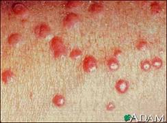
3. Tendon xanthoma: classically a slowly appearing nodule on the extensor tendons of hands and feet.


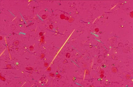
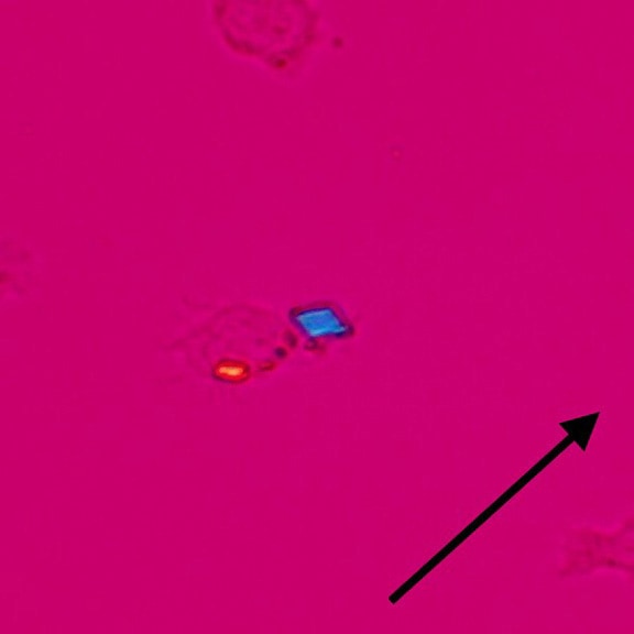
4. Lipemia retinalis: Very high levels of triglycerides (>400 in the USA, >40 in the rest of the world). You can see a milky colouration to the retinal vessels.


A neat link from NEJM on lipemia retinalis over here.

Thursday, October 7, 2010

Rheumatoid Arthritis: Some hand findings

On inspection remember to look for "SEADS" - swelling, erythema, atrophy, deformities, skin changes.


Wrist:
  • Often has Radial deviation
  • You may see a 'dinner fork' deformity at the wrist

Hand:

  • Look for muscle atrophy in the interossious, thenar, and hypothenar regions
  • Look and feel for Tendon nodules, and contractures

Fingers:

  • Vasculitic nail changes: nail fold infarcts, periungual erythema
  • Ulnar deviation of MCP's
  • Swelling of the PIP's, the DIP's become involved later in the disease
  • Swan Neck deformities (see photo above)
  • Boutonniere deformitiy (see photo above)


Tuesday, October 5, 2010

Cranial Nerves III, IV, and VI


Interesting eye findings today:


Cranial Nerve III
  • All extra ocular movements except those from the Superior Oblique (CN IV) and Lateral Rectus (CN VI).
  • Innervates the Levator Palpebrae Superioris: this keeps your eyelid open. Many CN III lesions will result in ptosis.
  • Efferent limb of the pupillary light reflex. Remember to look for a "direct" and then a "consensual" response to light.
  • A Quick CN III Tip: Pupillary constriction is controlled by the outer fibers of CN III, and ocular movements/upper eyelids are controlled by the inner fibers. So lesions that compress the outer CN III fibers (eg. lesions like Posterior Communicating Artery Aneurysms) present initially with dilated pupils, while lesions affecting the inner portions of CN III (like infarction in diabetes) present with ptosis and a "down and out" position of the eye that spares the pupil.

Cranial Nerve IV

  • Innervates the Superior Oblique muscle, which moves the eye "down and in". Lesions here result in difficulty looking down. Patients have trouble walking downs stairs.

Cranial Nerve VI

  • Innervates the Lateral Rectus muscles, which moves the eye laterally.

Thursday, September 30, 2010

Tophi and Gout

Today we saw some pretty empressive gouty tophi - small joints on the hands were involved, as well as an impressive tophus on the elbow (this picture is from wikipedia, not our patient). Tophi are just uric acid crystals deposited in the tissues, and is evident usually after several years of gout. Remember to look for tophi on the joints, but also in the helix/antihelix of the ear (see picture below).


Gout:
  • Clinical: Typically presents with acute monoarthritis, occasionally a few joints involved. Red, hot, swollen, tender. Often first MTP, ankle, knee.
  • Evaluation: Negatively birefringent crystals visualized, often intracellular. They look like this. These are monosodium urate crystals.
  • Risk factors: hyperuricemia (lymphoma, other causes of increased cell turnover), obesity, EtOH, medications like HCTZ, asa.
  • Acute Treatment: NSAIDS, intra-articular steroids, colchicine, systemic steroids if multiple joints involved.
  • Chronic treatment: Allopurinol if multiple attacks per year (usually >2).
Pseudo Gout:
  • Rhomboid crystals, occasionally inside macrophages. They look like this. These are calcium pyrophosphate crystals.
  • Often in elderly patients with multiple medical problems.
  • Often polyarticular, and may affect wrists, shoulders.

Tophi on the ear

Tuesday, September 28, 2010

Knee Effusion

-Tunisia?
-Two Knees, Yeah!
-Two Knees, Huh?

Okay...terrible...there's plenty more where that came from. Today we examined two knees. And a few knee problems - gout, prepatellar bursitis, and septic arthritis.

Is there an effusion?

1. Inspect: in the absence of an obvious effusion, have a look at the medial side of the knee - normally there is a 'dimple' here. Expose both sides and compare. Absence of this dimple is usually indicative of an effusion.

2. Palpate: There are a few ways to feel for an effusion....
  • Patellar Tap: push on the suprapatellar region to get fluid under the patella, then press firmly on the patella. You may feel it tap on the bone underneath. Kind of uncomfortable.
  • Ballotment: push on the suprapatellar region to get fluid under the patella. Then with your other hand, use your thumb and index finger to ballot the fluid back and forth.
  • "Milk" test: 'milk' (or 'sweep') fluid from the medial side up the knee and watch that medial dimple re-appear. Then sweep the fluid back down the lateral aspect of the knee and see the medial dimple bulge out again.
  • The Forgotten 4th test: Not sure what to call this one. I learned from Dr. Mary Bell, an incredible Rheumatologist. With one hand, place your index finger and thumbs on either side of the patella. Then with your index finger of your other hand, gently press on the suprapatellar area. In the presence of an effusion, you will feel pressure on your thumb and forefinger.

Let me know if you need this explained again at the bedside - happy to show you at any time.

3. Links:


Thursday, September 23, 2010

Chest findings...."just tap it in...tap it in..."


Today we did a lot of percussion and auscultation....and a bit of tactile vocal fremitus. The summary below is very important - you can predict what you will see on chest x-ray with your history and physical exam >90% of the time. Click on it to enlarge. Here is a good link to the complete chest physical exam.