Thursday, September 30, 2010

Tophi and Gout

Today we saw some pretty empressive gouty tophi - small joints on the hands were involved, as well as an impressive tophus on the elbow (this picture is from wikipedia, not our patient). Tophi are just uric acid crystals deposited in the tissues, and is evident usually after several years of gout. Remember to look for tophi on the joints, but also in the helix/antihelix of the ear (see picture below).


Gout:
  • Clinical: Typically presents with acute monoarthritis, occasionally a few joints involved. Red, hot, swollen, tender. Often first MTP, ankle, knee.
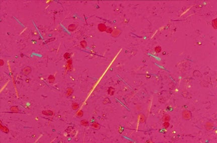
  • Evaluation: Negatively birefringent crystals visualized, often intracellular. They look like this. These are monosodium urate crystals.
  • Risk factors: hyperuricemia (lymphoma, other causes of increased cell turnover), obesity, EtOH, medications like HCTZ, asa.
  • Acute Treatment: NSAIDS, intra-articular steroids, colchicine, systemic steroids if multiple joints involved.
  • Chronic treatment: Allopurinol if multiple attacks per year (usually >2).
Pseudo Gout:
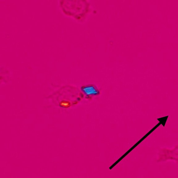
  • Rhomboid crystals, occasionally inside macrophages. They look like this. These are calcium pyrophosphate crystals.
  • Often in elderly patients with multiple medical problems.
  • Often polyarticular, and may affect wrists, shoulders.

Tophi on the ear

Tuesday, September 28, 2010

Knee Effusion

-Tunisia?
-Two Knees, Yeah!
-Two Knees, Huh?

Okay...terrible...there's plenty more where that came from. Today we examined two knees. And a few knee problems - gout, prepatellar bursitis, and septic arthritis.

Is there an effusion?

1. Inspect: in the absence of an obvious effusion, have a look at the medial side of the knee - normally there is a 'dimple' here. Expose both sides and compare. Absence of this dimple is usually indicative of an effusion.

2. Palpate: There are a few ways to feel for an effusion....
  • Patellar Tap: push on the suprapatellar region to get fluid under the patella, then press firmly on the patella. You may feel it tap on the bone underneath. Kind of uncomfortable.
  • Ballotment: push on the suprapatellar region to get fluid under the patella. Then with your other hand, use your thumb and index finger to ballot the fluid back and forth.
  • "Milk" test: 'milk' (or 'sweep') fluid from the medial side up the knee and watch that medial dimple re-appear. Then sweep the fluid back down the lateral aspect of the knee and see the medial dimple bulge out again.
  • The Forgotten 4th test: Not sure what to call this one. I learned from Dr. Mary Bell, an incredible Rheumatologist. With one hand, place your index finger and thumbs on either side of the patella. Then with your index finger of your other hand, gently press on the suprapatellar area. In the presence of an effusion, you will feel pressure on your thumb and forefinger.

Let me know if you need this explained again at the bedside - happy to show you at any time.

3. Links:


Thursday, September 23, 2010

Chest findings...."just tap it in...tap it in..."


Today we did a lot of percussion and auscultation....and a bit of tactile vocal fremitus. The summary below is very important - you can predict what you will see on chest x-ray with your history and physical exam >90% of the time. Click on it to enlarge. Here is a good link to the complete chest physical exam.


Tuesday, September 21, 2010

Splenomegaly

What does this picture have to do with splenomegaly? Send me an email - if you're right, I'll buy you a coke.

The spleen is the 3rd most underappreciated internal organ.

If you need a reminder on how to perform any of the maneuvers below, send me an email/page and we can go through it.

Does my patient have splenomegaly? Tough to say... this is not the most reliable exam, however if your pre-test probability is somewhat high (>10%), then the exam can be quite helpful.

Castell's Sign: The patient is supine. Locate the intersection between the left anterior axillary line (*note* not mid axillary line) and the last intercostal space. Perucuss. It should be resonant. When your patient breaths in and holds, this should stay resonant. If you hear dullness then voila - probably splenomegaly(Sensitivity 82%; Specificity 83%).

Palpation: Either use your hand, or the patients hand for counter-pressure in the left flank. Palpate from the RLQ diagonally up, inching incrementally as the patient exhales. Try rolling the patient into the right lateral decubitus if you do not feel anything and repeat your palpation(Sensitivity ~60%; Specificity ~90%). Feel for the splenic notch.

Middleton's Hooking Maneuver: Approach the patient from the left side, with the patient supine and their fist under the left costovertebral angle, hook your fingers under the left costal margin. Ask the patient to breath in, and feel for the spleen tip (sensitivity ~60%; Specificity ~90%).

Others: Nixon's percussion method is helpful, but Traube's space is pretty useless. I can show you these whenever you like.

Here is a link to the sentinel JAMA article on splenomegaly.

Thursday, September 16, 2010

Stevens-Johnson Syndrome

Stevens-Johnson Syndrome and Toxic Epidermal Necrolysis are about as bad a drug reaction as you can get. These are basically the same condition, with TEN being on the more serious side of the spectrum compared to SJS

SJS: Less than 10% of body surface area is affected. Mucous membranes virtually always involved. macules which are erythematous or purpuric morph into epidermal necrosis and sloughing.

TEN: See above, except >30% of the body is involved. Diffuse erythema is also seen in this condition.

Both: Associated with systemic symptoms like fever, malaise, anorexia. Bullae may form on the skin prior to sloughing.

What causes it? Mostly in response to medications. Classic drugs include allopurinol, sulfa medications, penicillins, anti-seizure drugs (dilantin, carbamazepine), and some NSAIDS.

How do your manage these cases? 1. early recognition, 2. discontinuation of offending agent, 3. supportive care, 4. wound care management, including eyes and mouth, 5. monitor for fluid/electrolyte disturbances, 6. debridement. The use of IVIG or steroids is controversial. Many patients require specialist care in a burn unit.

How does the picture above relate to this topic? send me an email...if you're right I'll give you a prize.

Tuesday, September 14, 2010

Terry's Nails and Lindsay's Nails

Today we looked at nails a bit more closely. We examined the white part of the nail, termed the Lunula, which is Latin for 'little moon'. This normally occupies a very small proportion of your nail.


Lindsay's Nails: also termed "half-and-half" nails. As the name implies, the lunula extends to roughly 50% of the nail. It is classically described in chronic kidney disease.

Terry's Nails: The lunula extends to >75% of the nail. This is described in liver diseases, CHF, diabetes, and hyperthyroidism.

Thursday, September 9, 2010

Interstitial Lung Disease

Today we saw some really interesting cases including:

*Ascites: check out a previous post with evidence-based physical exam findings here.

*Clubbing: also discussed, with evidence-based exam findings at this link.

One patient we saw had terrible interstitial lung disease. These are confusing conditions. Here's an approach as to the underlying etiology:

Systemic Diseases: ILD is found commonly in Sarcoidosis and Collegen Vascular Diseases - the prototype being Scleroderma. Remember, ILD is not so common in SLE or Rheumatoid arthritis.

Exposures: these are classically divided into organic and inorganic exposures:

  1. Organic: aka "Hypersensitiviy Pneumonitis". Think about bird fanciers lung, cheese washers lung, farmers lung, and many many more.
  2. Inorganic: aka "Pneumoconiosis". This includes dust from asbestos, silica, and coal.
Drugs: Exposure to alkylating chemotherapeutic agents (eg. bleomycin), but also commonly used drugs like amiodarone, methotrexate, nitrofurantoin, and sulfa medications are also implicated in ILD.

Idiopathic: This is a strange category with bizarre acronyms. It doesn't help that there are old and new acronyms, and that both are used frequently. The diagnosis is usually based on histology.
  1. Usual Interstitial Pneumonia (UIP), also known as Interstitial Pulmonary Fibrosis (IPF) - this is perhaps the most common idiopathic ILD.
  2. Acute Interstitial Pneumonia (AIP), also known as the 'Hamman-Rich Syndrome'. Just like IPF above, but more rapid.
  3. Cryptogenic Organising Pneumonia (COP), but if the underlying etiology is known, you will hear it called Bronchiolitis Obliterans and Organising Pneumonia (BOOP). There is lots of granulation tissue in the small airways here.
  4. Others: Desquamative Interstitial Pneumonia (DIP), Lymphocytic Interstitial Pneumonia (LIP), Non-specific Interstitial Pneumonia (NSIP)....oy vey...
Other Rare Causes of ILD: Alveolar hemorrhage, Pulmonary Alveolar Proteinosis, and my personal favorite, Pulmonary Infiltrates with Eosinophilia (PIE). mmmmmh.....pie. And Lymphangioleiomyomatosis (LAM).

Mimickers of ILD: Pneumocystis Carini Pneumonia, Lymphangitic carcinomatosis, and once in a while, good ol' congestive heart failure.

Links: A good review of Idiopathic Pulmonary Fibrosis can be found at this link.

Tuesday, September 7, 2010

Clubbing


Common causes of Clubbing:
  • Gastrointestinal: liver disease, inflammatory bowel diseases, celiac disease, malignancy
  • Pulmonary: malignancy, suppurative (empyema, abscess), Cystic Fibrosis, TB, bronchiectasis, many fibrosing conditions (like IPF), AVM's
  • Cardiac: endocarditis, cyanotic heart diseases
  • Other: hyperthyroidism, alpha 1 antitrypsin, sarcoid, lymphoma

Physical Examination for clubbing:

  • Look for changes in the nail shape, like increased PA and lateral diameter, and a "bird-beaking" or "drumstick" appearance to the nail.
  • Palpate a spongy texture to the nailbed.
  • "Phalangeal Depth Ratio": See the picture below in Frame C. Normally the finger height at the DIP should be greater than at the distal phalanx. In clubbing, the height at the distal phalanx is greater than the at the DIP.
  • Angles: See the picture below in Frame B: the Profile angle (also known as Lovibonds angle) is increased greater than 180 degrees in clubbed individuals - this is angle ABC below; and the Hyponychial angle is greater than 190 degrees in clubbing as well - this is angle ABD below.
  • Shamroth Sign: See the picture below. This is an indirect measurement of the Profile angle (aka Lovibonds angle). When you put two fingers together at the terminal phalanx, you should see a diamond shape. This is lost in clubbed individuals.

What is the evidence for all this?

  • JAMA has a good article in their Rational Clinical Exam Series at this link.
  • There is a recent paper specific to the utility of Shamroth's sign in JAMA here as well.
  • Bottom line: the Phalangeal Depth Ratio and Shamroth Sign are the more useful findings.


(Image is from the JAMA Rational Clinical Exam series; 2001, 286, 341-47 at this link)

Thursday, September 2, 2010

Edema

Today we saw of case of severe non-pitting edema, and a case of what appeared to be cellulitis, but in fact was radiation-induced skin changes.

An approach to Edema is below.

Here is a link to diseases which may masquerade as cellulitis from the Annals of Internal Medicine.


Edema is a common finding in clinics and on the wards. It is usually found in dependent areas like the ankles and legs, but may be seen in the sacrum of supine individuals.

Pitting vs Non-Pitting: to have 'pitting' edema, a dent is left in the skin after pressure is applied over a bony prominence. How long do you have to press? Some say 5 seconds, however many experienced clinicians agree that 15-20 seconds is more appropriate.
  • Non-Pitting: Suggests lymphedema, or rarely hypothyroidism (pretebial myxedema).
An Approach to Bilateral Leg Edema:
  • Cardiogenic: think about right-sided heart failure or constrictive pericarditis
  • Venous Obstruction: should be above the level of iliac veins, or bilateral distal venous thrombus
  • Hepatic: when the synthetic function declines, decreased protein production like in late cirrhosis
  • Other GI: protein losing enteropathies, malnutrition
  • Kidney: nephrotic syndrome
  • Endocrine: Cushing's syndrome - high cortisol levels increase renal sodium absorption. Also remember hypothyroidism (pretibial myxedema)
  • Drugs: calcium channel blockers, OCP, corticosteroids, minoxidil
  • Pregnancy: an planned (or unplanned?) mass pressing on IVC

Unilateral Edema: this may be caused by

  • Venous disease: such as DVT, thrombophlebitis, damaged venous valves after infection or clot.
  • Lymphatic disease or disruption: often lymph nodes are removed during surgery (eg for breast cancer). Infection is a common cause of LN damage, such as in recurrent cellulitis or lymphatic filariasis. You may see unilateral edema from LN disease with certain malignancies or radiation-induced damage as well.