Tuesday, August 31, 2010

Aortic Stenosis Redux


(rainbow trout pictured left)

Today we saw a case of severe aortic stenosis (valve area 0.6 cm).


We have discussed AS in detail before, and you can check out this link for more information on the physical exam.

Can you detect moderate to severe AS at the bedside? Absolutely. Some smart people from Toronto came up with a very helpful clinical prediction rule. The published paper from JGIM is at this link.

This is how it works:


  • If there is no murmur over the right clavicle, moderate to severe AS is essentially ruled out.
  • If there is a murmur radiating over the right clavicle with 0-2 associated findings, then the Likelihood Ratio for moderate to severe AS is 1.8, and in this study reflected about a 20% chance.
  • If there is a murmur radiating to the right carotid with 3-4 associated findings, then moderate to severe AS is essentially ruled in (Likelihood ratio of 40)

So what are the "Associated Findings"?

  • Reduced S2
  • Reduced carotid volume
  • Slow carotid upstroke
  • Murmur loudest in the 2nd right intercostal space

Thursday, August 26, 2010

Guillain-Barré syndrome


Guillain-Barre Syndrome (GBS) is an umbrella term to describe a few syndromes of immune-mediated demyelinating polyneuropathy.


This is commonly manifested by:


  • Motor findings: typically weakness starts in the legs. It may ascend rather quickly and affect respiratory muscles. Facial muscles are also commonly involved. Reflexes are severely diminished to absent.

  • Sensory changes: found usually in extremities. Typically mild decreases to light touch, pain, temperature. Pain in the lower extremities and back are also a common feature.

  • Autonomic findings: patients may present with tachycardia, bradycardia, hypotension, urinary retention.


These symptoms may start abruptly and develop over a period of hours, days, to a couple of weeks.

A related variant: the Miller-Fischer Syndrome: This is under the same umbrella as GBS and is manifested by 1. Ophthalmoplegia, 2. Ataxia, 3. Areflexia. Patients will also usually have peripheral weakness.

Diagnosis: You will hear the term "Albuminocytologic Dissociation". This means that in the CSF there are elevated protein levels with normal WBC counts. This is a classic feature of GBS. Nerve conduction studies will also help clinch the diagnosis. MRI will often show enhancement of nerve roots.

Treatment: with plasma exchange or IVIG therapy. This is to eliminate or incapacitate auto-antibodies to schwann cells. Patients must be watched closely for possible respiratory muscle compromise. Roughly 80% will make a full recovery.

Risk Factors: Autoantibodies may form after exposure/infection:

  • Campylobacter
  • EBV/CMV
  • HIV
  • Hodgkin's Disease
  • Influenza vaccine? If real, likely a tiny risk.

A few good links:

Tuesday, August 24, 2010

Mitral Regurgitation

Mitral Regurgitation is pretty common. when you hear this murmur and are trying to determine the underlying cause, think about the individual components of the mitral valve and particular disease states which might affect them. Let's start at the annulus and work our way down....


  1. Annulus: This may be dilated from cardiomyopathies, or calcified in diseases like rheumatic fever or chronic renal insufficiency.
  2. Leaflets: The mitral leaflets can fail in a number of disease states including infectious endocarditis (acute or chronic), rheumatic fever, autoimmune diseases (SLE, scleroderma), myxomatous degeneration (MVP), Connective Tissue Diseases like Marfan's syndrome, or with congenital anomalies.
  3. Chordae: These can be damaged or rupture under ischemic, infected, or traumatic conditions, and in rheumatic heart disease.
  4. Papillary Muscles: These muscles can rupture after trauma or infarct. They become 'dysfunctional' in ischemic conditions or when the LV becomes dilated (myopathy or aneurysm). Papillary muscles can also become infected, and rarely can be infiltrated with amyloid deposits or granuloma (eg. sarcoid).
On Exam:

  • Inspection/Palpation: Apical impulse may be displaced to the left and is brisk and hyperdynamic.

  • Heart Sounds: S1 may be soft, S2 may be widely split from early A2 closure. You may hear a loud P2 if pulmonary hypertension is present. S3 can be heard in volume overloaded states.

  • Murmur: This is a holosystolic murmur that starts right after S2 (and may even obscure it). It is high-pitched, loudest at the apex, and radiates to the axilla. There is minimal respiratory variation - this may help you distinguish it from Tricuspid Regurgitation.

  • Special Tests: The murmur is accentuated with maneuvers that increase afterload (eg. bilateral isometric hand grip or by transient arterial occlusion with a blood pressure cuff). The murmur will be diminished by decreasing preload - like going from a crouching position to standing, or with valsalva.

Good Links:

Thursday, August 19, 2010

Aortic Insufficiency

(a leaky valve, pictured left)


Aortic Insufficiency: probably the most eponymous physical exam scenario....


  • Pathophisiology: increased stroke volume results in an abrupt distention of peripheral arteries and elevation in blood pressure. This is followed by regurgitation through the valve back into the LV, causing a rapid fall in pressure with quick collapse for the arteries and a low diastolic pressure. This accounts for the majority of physical exam findings.

  • Inspection of the Precordium: the apex may be displaced laterally or inferiorly.

  • Palpation: displaced cardiac apex, with a hyperdynamic pulse. You may feel a thrill near the sternal notch.

  • Heart Sounds: S1 may be soft secondary to a longer PR interval. An S3 will be heard in volume overloaded states.

  • Murmur: typically a high-pitched early diastolic murmur, decrescendo in nature. It is best heard with a patient sitting up and leaning forward at end exhalation. Classic teaching suggests that if the murmur is loudest at the Right Sternal Boarder it represents aortic root disease, and if the murmur is loudest at the Left Sternal Border it represents aortic valve disease.

  • Other Murmurs you may hear: A systolic murmur at the base that may mimic aortic stenosis - this is usually just a murmur from the high volume and flow of blood through the aortic valve during systole. Why so much during systole? Because a large fraction is regurgitationg back into the LV during during diastole. Also, an Austin-Flint Murmur may be heard. As Dr. Davis pointed out, this is a diastolic murmur heard at the apex of the heart that may mimic mitral stenosis.

Etiology of Aortic Insufficiency:

  • Aortic Valvular Disease: Rheumatic heart disease, endocarditis, bicuspid aortic valve, myxomatous degeneration, rheumatoid arthritis, Marfan's, ankylosing spondylitis.
  • Aortic Root Disease: HTN, dissecting aortic aneurysm, aortitis (from syphilis, ankylosing spondylitis, IBD, reactive arthritis), rare connective tissue diseases like Ehlers Danlose syndrome.

What's with all the eponyms? These are cool to see, but largely useless as they are not sensitive or specific, and are mostly related to the wide pulse pressure.

  • DeMusset's sign: head bobbing with each heart beat.
  • Becker's sign: visible pulsations in the retinal arterioles.
  • Mueller's sign: systolic pulsations of the uvula.
  • Quincke's sign: capillary pulsations in the finger tips.
  • Rosenbach's sign: systolic pulsations in the liver.
  • Gerhard's sign: systolic pulsations in the spleen. Usually you need an enlarged spleen to feel.
  • Duroziez's sign: a systolic and diastolic bruit heard over the femoral artery when it is partially compressed. Note that when the distal part of your stethoscope is pressed down, the diastolic component will be accentuated.
  • Pistol Shot sound: heard over the femoral artery.
  • Hill's sign: when BP at the popliteal region is greater than the brachial BP by more than 20 mmHg.
  • Water-hammer pulse aka Corrigan's sign: with a patient lying down, press down to obliterate the radial pulse. Then slowly raise their arm up and voila! The pulse becomes palpable again with the same amount of applied pressure.

So, is there any evidence for all this?

A little bit. Some smart folks in Toronto wrote a good JAMA article that you can read at this link. AI can be ruled out with the absence of an early diastolic murmur, and ruled in with the presence of an early diastolic murmur - the caveat? These murmurs were heard by cardiologists. Perhaps mere mortals like ourselves will hear it too.

Tuesday, August 17, 2010

Dermatomyositis


(above left: heliotrope rash, above right: Gottron's papules)

Classic skin manifestations of Dermatomyositis include:
  • Gottron's papules: symmetric, erythematous and scaly appearance over the MCPs/PIPs/DIPs. You may also see this on the elbows.
  • Heliotrope rash: classically described as a 'violaceous' rash on the eyelids.
  • Shawl Sign: Imagine your patient is wearing a shawl - there is often erythema in a 'V' shaped distribution over where a shawl would be worn.

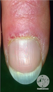
  • More Subtle Signs: You may hear the term "Mechanics Hands" which refers to a rugged and cracked appearance of the lateral aspect of the fingers, usually at the end - and as the name implies, this is what the hands of a manual labourer may appear like. Another finding is periungual edema, and dilated periungual vessels.

*A good way to test for proximal muscle weakness in this condition is to have a person sit in a chair with their arms folded across their chest, and have them attempt to stand up.

*Remember that many patients with Dermatomyositis have an underlying malignancy, and the diagnosis of Dermatomyositis should prompt an age-appropriate and risk-factor-appropriate malignancy screen. This link shows some new data relating the two conditions.

(periungual vascular changes pictured left)

Thursday, August 12, 2010

Systemic Sclerosis

(telangiectasia pictured left)

Systemic Sclerosis (aka scleroderma) is an autoimmune condition affecting the skin and internal organs. There are 2 forms of Systemic Sclerosis outlined below - but there are also varieties limited to the skin :


        • Diffuse Cutaneous Systemic Sclerosis: as the name implies, there are diffuse skin manifestations, including the torso, arms, legs, face.

        • Limited Cutaneous Systemic Sclerosis. skin involvement is usually limited to the hands, feet and face. We often talk about the CREST syndrome - this is seen in the Limited Cutaneous Systemic Sclerosis.


        CREST Syndrome:

        • Calcinosis: calcium deposits in soft tissue.
        • Raynaud's phenomenon: with vasospasm the digits first turn white, then blue, then a red hyperemic reaction.
        • Esophageal dysmotility.
        • Sclerodactyly: tightening of the skin around fingers and toes, and eventual loss of subcutaneous tissue.
        • Telangiectasia: dilated blood vessels which blanch with pressure.


        Here is a good link to a review on Scleroderma.


        (sclerodactyly pictured left)


        Tuesday, August 10, 2010

        Bullous Pemphigoid


        Bullous Pemphigoid pictured left, and a great review of bullous skin diseases from the American Family Physician at this link.


        Bullous Pemphigoid: A rare, autoimmune blistering disease that is seen primarily in the elderly. It is from IgG autoantibodies against the basement membrane.


        How do you distinguish Bullous Pemphigoid from Pemphigus Vulgaris?
        • Bullous Pemphigoid: tense bullae, pruritic, less oral involvement, and Nikolsky's sign usually negative.
        • Pemphigus Vulgaris: flaccid and ruptured bullae, non-pruritic, oral involvement very common, Nikolsky's sign positive.

        A diagnosis is confirmed via biopsy and staining for the IgG auto-antibodies, and treatment usually involves corticosteroids. Bullous Pemphigoid typically has a good prognosis.

        Thursday, August 5, 2010

        Pericardial Rubs


        Today we heard a pericardial rub.

        So what is the first thing to do when you hear one of these? Go find your friend and get them to listen to it too. Pericardial rubs are notoriously transient, and when your attending looks at you with disdain the next day as he or she hears normal heart sounds, you've got your friend to back you up that it indeed was present at 3:00 am.

        What does it sound like? "leather on leather".....kind of like in that movie you saw but won't admit to anybody.

        Where is it most commonly heard? At the left sternal boarder.

        Phases to the rub? you may hear three components of the rub, correlating with 1. atrial systole, 2. ventricular systole, and 3. ventricular diastole. Just over 50% of rubs have 3 phases, and you can hear 2 phases in another 30%.

        When do you hear this? A common finding in pericarditis. You will see it with the following conditions:
        • Idiopathic: common - likely viral.
        • Infectious: Viral (common), TB (also common) bacterial (uncommon), fungal
        • Autoimmune: SLE, rheumatic fever, drug induced lupus
        • Metabolic: Uremia
        • Malignant: eg lung or breast cancer with metastases, lymphoma
        • Radiation
        • Post MI: Dressler's syndrome
        • Structural: aortic dissection with blood leaking into pericardium, post pericardiotomy, post cardiac surgery, post trauma

        Here is a very good review of pericarditis.

        Tuesday, August 3, 2010

        Ascites


        Today we saw an interesting case of ascites. A few things to remember:

        History: Ask about recent weight gain and increasing abdominal girth. Also remember to ask about ankle edema. Is there a history of liver disease or risk factors for liver disease?

        Inspection: Best from the foot of the bed - have a look for bulging flanks, dilated abdominal veins and umbilical hernia.

        Percussion: Percuss for flank dullness, then for shifting dullness. HINT - it is easier to perform this manoeuvre when you percuss from medial to lateral, and roll the patient AWAY from you.

        Fluid Wave: place the patients hand in the middle of the abdomen to block the 'adipose wave,' then flick or tap pretty sharply on one side while feeling for the wave on the contralateral side. Some of us are "tappers" while others are "flickers". It's like the Red Sox versus the Yankees. What unites us all is that we block the fluid wave, and firmly (yet politely) elicit this finding.

        Other: Look for pitting edema at the ankles, and for stigmata of liver disease.

        Okay...great. So what is the evidence for all this? Does it work? I heard there is something called the "Puddle Sign".... yeah.... don't do that one.

        There is a good JAMA article on ascites you can read at this link, and summarized below:

        Sensitive tests: Ankle swelling, increasing abdominal girth, bulging flanks, and flank dullness.
        Specific tests: Fluid Wave is the best